Friday, May 4, 2007

Acute Pancreatitis Signs and Symptoms

These are signs and symptoms of Acute pancreatitis: Acute pancreatitis comes on suddenly, usually with mild to severe pain in your upper abdomen that may radiate to your back and occasionally to your chest. The pain may be nearly constant for hours or even days and is likely to become worse when you drink alcohol or eat. Bending forward or curling into a fetal position may provide temporary relief.


Other signs and symptoms of acute pancreatitis include:
  1. Nausea;
  2. Vomiting;
  3. Fever;
  4. Rapid pulse;
  5. Swollen, tender abdomen.
In severe cases:
  1. Dehydration;
  2. Low blood pressure;
  3. Internal bleeding; and
  4. Shock.
You may have repeated episodes of acute pancreatitis and recover fully from each one. Nevertheless, every attack is a serious illness that can damage your pancreas and cause life-threatening complications.

To view information on another disease, click on Pancreatitis SOD Library.!

Pancreatitis SOD Library


..


Chronic Pancreatitis Signs and Symptoms

These are signs and symptoms of Chronic pancreatitis: Ongoing damage to your pancreas can lead to a chronic condition that destroys the pancreas and nearby tissues, although it may be years before signs and symptoms appear. A few people with chronic pancreatitis never experience discomfort, but most have intermittent bouts of abdominal pain that can be severe. The pain may last anywhere from a few hours to weeks or even years. Drinking alcohol, smoking tobacco, or eating often makes symptoms worse.
In addition to pain, chronic pancreatitis can cause:
  1. Nausea;
  2. Vomiting;
  3. Fever;
  4. Bloating and gas;
  5. Weight loss, even when your appetite and eating habits are normal;
  6. Steatorrhea: Oily, malodorous stools resulting from poor digestion and malabsorption of nutrients, particularly fats;
  7. Diabetes;
  8. Sometimes, jaundice.

To view information on another disease, click on Pancreatitis SOD Library.!

Pancreatitis SOD Library


..


Causes

Causes


  • Most common causes: A common mnemonic for the causes of pancreatitis is:
    ........1. I -
    idiopathic
    ........2. G -
    gallstone. Gallstones that travel down the common bile duct and which subsequently get stuck in the Ampulla of Vater can cause obstruction in the outflow of pancreatic juices from the pancreas into the duodenum. The backflow of these digestive juices causes lysis (dissolving) of pancreatic cells and subsequent pancreatitis.
    ........3. E -
    ethanol (alcohol)
    ........4. T -
    trauma
    ........5. S -
    steroids
    ........6. M-
    mumps (paramyxovirus) and other viruses (Epstein-Barr virus, Cytomegalovirus)
    ........7. A -
    autoimmune disease (Polyarteritis nodosa, Systemic lupus erythematosus)
    ........8. S -
    scorpion sting - Tityus Trinitatis - Trinidad/ snake bite
    ........9. H -
    hypercalcemia, hyperlipidemia/hypertriglyceridemia and hypothermia
    ......10. E -
    ERCP (Endoscopic Retrograde Cholangio-Pancreatography - a form of endoscopy)
    .......11. D-
    .
    Drugs
    (SAND) And Duodenal Ulcers
    .................. a. S -Steroids and Sulphonamides
    ...................b. A- Azathiopine
    ...................c. N - NSAIDS
    ...................d. D - Diuretics such as:
    ..............................1. Furosemide
    ..............................2. Thiazides
    ..............................3. Didanosine
  • Less common causes
    .......,1. pancreas divisum
    ........2. long common duct
    ........3. carcinoma of the head of pancreas, and other cancer
    ........4. ascaris blocking pancreatic outflow
    ........5. ischemia from bypass surgery
    ........6. fatty necrosis
    ........7. pregnancy
    ........8. infections other than mumps, including varicella zoster
    ........9. repeated marathon running.
  • Causes by demographic:
    ........1. Western countries - chronic alcoholism and gallstones accounting for more than 85% of all cases
    ........2. Eastern countries - gallstones
    ........3. Children - trauma
    ........4. Adolescents and young adults - mumps

To view information on another disease, click on Pancreatitis SOD Library.!

Pancreatitis SOD Library


..


Screening and Diagnosis

Screening and diagnosis of Pancreatitis: Because diagnosing pancreatitis can be difficult, you're likely to have several tests to help pinpoint the problem. The type of test may depend on whether your pancreatitis is acute or chronic.
...
Acute pancreatitis: If your doctor suspects that you have acute pancreatitis, a sample of your blood may be analyzed for abnormalities such as:
  1. Elevated levels of the pancreatic enzymes, amylase and lipase;
  2. Elevated white blood cell count;
  3. Elevated liver enzymes and bilirubin, a substance that results from breakdown of red blood cells;
  4. High blood sugar (hyperglycemia);
  5. Low calcium level — high calcium levels can cause pancreatitis, but low levels of calcium in the blood, called hypocalcemia, are a common result;
Because laboratory tests can't confirm a diagnosis of acute pancreatitis, your doctor may request an ultrasound or computerized tomography (CT) scan of your abdomen to examine your pancreas and to check for gallstones, a duct problem, or destruction of the gland. You may also have X-rays of your abdomen and chest to rule out other reasons for your symptoms.
..
Diagnosing Chronic pancreatitis:
Once there is damage in the pancreas, it is considered Chronic Pancreatitis. Chronic Pancreatitis is an ongoing progressive disease. There is no cure, but there is hope in research.
..
Diagnosing chronic pancreatitis can be challenging because some tests may yield normal results, even though you have the disease. It can also be difficult to distinguish acute from chronic pancreatitis. Even so, certain tests can help rule out other problems and aid in the diagnosis. These include:
  • Blood tests. These tests can identify abnormalities associated with chronic pancreatitis and help rule out acute inflammation.
  • Stool test. This measures the fat content in your feces. Chronic pancreatitis often causes excess fat in your stool because the fat isn't digested and absorbed normally by your small intestine.
  • Ultrasound. In standard (external) ultrasound, a wand-like device (transducer) is placed on your body. It emits inaudible sound waves that are reflected to the transducer and then translated into a moving image by a computer. Endoscopic ultrasound may provide images of your pancreas and bile and pancreatic ducts that are superior to those produced by standard ultrasound.
  • EUS. In endoscopic ultrasound, your doctor uses a thin, flexible tube with a light (endoscope) to thread a small ultrasound device through your stomach. The device then generates a detailed image on a computer screen.
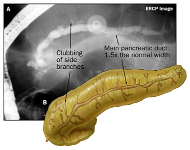
  • ERCP. X-ray of bile and pancreatic ducts. In a procedure called endoscopic retrograde cholangiopancreatography, your doctor gently threads an endoscope down your throat and through your stomach to the opening of the bile and pancreatic ducts in your duodenum. A dye passed through a thin, flexible tube (catheter) inside the endoscope allows for X-ray images of the ducts.

..

Fig. 1. Pancreas with no dialation of Main Pancreatic Duct and no clubbing of side branches.
....


Fig. 2. Pancreas with main pancreatic duct dialated to 1.5x normal width and some clubbing of side branches.

..

Fig. 3. Pancreas with main pancreatic duct dialated to > 1.5x normal width and clubbing of side branches.

...

  • Pancreatic function test. If you've lost weight or your doctor suspects a malabsorption problem, you may have a pancreatic function test. Several tests exist, but all measure the ability of your pancreas to secrete enzymes or other substances necessary for digestion.
  • SSMRC. MRCP With Secretion Stimulation. IV injection of synthetic secretin, a gut hormone, causes a rapid outpouring of bicarbonate-rich fluid from the exocrine pancreas. SSMRCP provides significantly improved visualization not only of the main PD, but also of its side branches, when compared to nonstimulated imaging.
  • You may need additional tests if your doctor is concerned about the possibility of other diseases, such as pancreatic cancer. Chronic pancreatitis puts you at a slightly higher risk of pancreatic cancer.

Complications: Severe cases of acute pancreatitis may lead to a number of complications:
  1. Infection. A damaged pancreas may become infected with bacteria that spread from the small intestine into the pancreas. Signs of infection include fever, an elevated white blood cell count and, in severe cases, organ failure. A fluid sample from the pancreas may be tested for bacterial infection. Pancreatic infections can be fatal without intensive treatment, including drainage or surgery to remove the infected tissue. Sometimes multiple operations are necessary.
  2. Pseudocysts. These are collections of pancreatic fluid and sometimes tissue debris that form within the pancreas or in an obstructed duct. If the cyst is small, no special care may be necessary, but large, infected or bleeding pseudocysts require immediate treatment.
  3. Abscess. This is a collection of pus in or near your pancreas that may develop about four to six weeks after the onset of acute pancreatitis. Treatment involves drainage of the abscess by catheter or surgery.
  4. Respiratory failure. Chemical changes in your body can affect your lung function, causing the level of oxygen in your blood to fall to dangerously low levels.
  5. Shock. This life-threatening complication usually occurs when your blood pressure is so low your organs can't carry out their normal functions. Severe shock can cause death within minutes if left untreated.
The complications common to acute pancreatitis can also occur in the chronic form of the disease. In addition, chronic pancreatitis can lead to:
  1. Bleeding. Ongoing inflammation and damage to the blood vessels surrounding the pancreas can cause potentially fatal bleeding.
  2. Malnutrition and weight loss. Lack of digestive enzymes prevents your body from absorbing nutrients from food. The result is often unintended weight loss and malnutrition.
  3. Diabetes. Damage to insulin-producing cells can lead to diabetes, a disease that affects the way your body uses blood sugar.
  4. Drug dependency. Because medical treatments for severe pancreatic pain aren't always effective, people with pancreatitis may become dependent on pain medications.
  5. Pancreatic cancer. Long-term inflammation of the pancreas increases your risk of pancreatic cancer, one of the most serious of all malignancies.

..

    Why Pain?

    The role of mast cells in the pathogenesis of pain in chronic pancreatitis.

    BMC Gastroenterol. 2005; 5:8 (ISSN: 1471-230X)
    Hoogerwerf WA; Gondesen K; Xiao SY; Winston JH; Willis WD; Pasricha PJEnteric Neuromuscular Disorders and Pain Laboratory, Division of Gastroenterology and Hepatology, University of Texas Medical Branch, 301 University Boulevard, Galveston, TX 77555-0764, USA.
    wahooger@utmb.edu

    • BACKGROUND: The biological basis of pain in chronic pancreatitis is poorly understood. Mast cells have been implicated in the pathogenesis of pain in other conditions. We hypothesized that mast cells play a role in the pain of chronic pancreatitis.We examined the association of pain with mast cells in autopsy specimens of patients with painful chronic pancreatitis. We explored our hypothesis further using an experimental model of trinitrobenzene sulfonic acid (TNBS) -induced chronic pancreatitis in both wild type (WT) and mast cell deficient mice (MCDM).
    • METHODS: Archival tissues with histological diagnoses of chronic pancreatitis were identified and clinical records reviewed for presence or absence of reported pain in humans. Mast cells were counted.The presence of pain was assessed using von Frey Filaments (VFF) to measure abdominal withdrawal responses in both WT and MCDM mice with and without chronic pancreatitis.
    • RESULTS: Humans with painful chronic pancreatitis demonstrated a 3.5-fold increase in pancreatic mast cells as compared with those with painless chronic pancreatitis. WT mice with chronic pancreatitis were significantly more sensitive as assessed by VFF pain testing of the abdomen when compared with MCDM.
    • CONCLUSION: Humans with painful chronic pancreatitis have an increased number of pancreatic mast cells as compared with those with painless chronic pancreatitis. MCDM are less sensitive to mechanical stimulation of the abdomen after induction of chronic pancreatitis as compared with WT. Mast cells may play an important role in the pathogenesis of pain in chronic pancreatitis.

    To view information on another disease, click on Pancreatitis SOD Library.!

    Pancreatitis SOD Library


    ..


    Pancreatic Pseudocysts

    Pancreatic pseudocyst
    From Wikipedia, the free encyclopedia

    Classification and external resources
    ICD-10
    K86.3
    ICD-9
    577.2
    DiseasesDB
    9530
    MedlinePlus
    000272
    eMedicine
    med/2674 radio/576
    MeSH
    D010192

    A pancreatic pseudocyst is a circumscribed collection of fluid rich in pancreatic enzymes, blood, and necrotic tissue (tissue that has died), typically located in the lesser sac of the abdomen. Pancreatic pseudocysts are usually complications of pancreatitis, although in children they frequently occur following abdominal trauma. Pancreatic pseudocysts account for approximately 75% of all pancreatic masses. The prefix pseudo- (Greek for "false") distinguishes them from true cysts, which are lined by epithelium; pseudocysts are lined with granulation tissue.

    Pathophysiology
    Acute pancreatitis results amongst other things in the disruption of pancreatic parenchyma and the ductal system. This results in extravasation of pancreatic enzymes which in turn digest the adjoining tissues. This results in a collection of fluid containing pancreatic enzymes, hemolysed blood and necrotic debris around the pancreas. The lesser sac being a potential space, the fluid collects here preferentially. This is called an acute pancreatic collection. Some of these collections resolve on their own as the patient recovers from the acute episode. However, others become more organized and get walled-off within a thick wall of granulation tissue and fibrosis. This takes several weeks to occur and results in a pancreatic pseudocyst.

    Investigations
    The questions that need to be answered are:
    1. Where, how big and how many?
    2. Is there a communication with the pancreatic ductal system? Draining such a pseudocyst carries an increased risk of pancreatic fistula.

    The most useful imaging tools are:
    1. Ultrasonography - The role of ultrasonography in imaging the pancreas is limited by patient habitus, operator experience and the fact that the pancreas lies behind the stomach (and so a gas-filled stomach will obscure the pancreas).
    2. Computerized tomography - This is the gold standard for initial assessment and follow-up
    3. Magnetic resonance cholangiopancreatography (MRCP) - to establish the relationship of the pseudocyst to the pancreatic ducts

    Treatment
    A small pseudocyst that is not causing any symptoms may be managed conservatively. However, a large proportion of them will need some form of treatment, The interventions available are:

    1. Endoscopic trans-gastric drainage
    2. Imaging guided percutaneous drainage
    3. Laparoscopic/open cystogastrostomy

    To view information on another disease, click on Pancreatitis SOD Library.!
    Pancreatitis SOD Library

    Treatment

    Treatment


    1. Relieving pain is the first step in treating chronic pancreatitis.
    2. The next step is to plan a diet that is high in carbohydrates and low in fat.
    3. A doctor may prescribe pancreatic enzymes to take with meals if the pancreas does not secrete enough of its own. The enzymes should be taken with every meal to help the body digest food and regain some weight.
    4. In some cases, surgery is needed to relieve pain. The surgery may involve draining an enlarged pancreatic duct or removing part of the pancreas.
    5. For fewer and milder attacks, people with pancreatitis must stop drinking alcohol, if they do drink, stop smoking tobacco, if they do smoke, stick to their prescribed diet, and take the proper medications.
    6. Sometimes insulin or other drugs are needed to control blood glucose.
    7. Complications will be treated if or as they arise (infections, pseudocysts, abscess, respiratory problems, shock, bleeding, malnutritian, weight loss, diabetes, drug dependency, pancreatic cancer.)

    Emergency Department Care: Most of the cases presenting to the ED are treated conservatively, and approximately 80% respond to such treatment:

    • Fluid resuscitation:
      • Monitor accurate intake/output and electrolyte balance of the patient.
        Crystalloids are used, but other infusions, such as packed red blood cells
        (PRBCs), are occasionally administered, particularly in the case of hemorrhagic pancreatitis.
      • Central lines and Swan-Ganz catheters are used in patients
        with severe fluid loss and very low blood pressure.
    • NPO: Patients should have nothing by mouth, and a nasogastric tube should be inserted to assure an empty stomach and to keep the GI system at rest.
    • **Begin parenteral nutrition if the prognosis is poor and if the patient is going to be kept in the hospital for more than 4 days.
    • Analgesics are used to relieve pain. Meperidine is preferred over morphine because of the greater spastic effect of the latter on the sphincter of Oddi.
    • Antibiotics are used in severe cases associated with septic shock or when the CT scan indicates that a phlegmon of the pancreas has evolved.
    • Other conditions, such as biliary pancreatitis associated with cholangitis, also need antibiotic coverage. The preferred antibiotics are the ones secreted by the biliary system, such as ampicillin and third generation cephalosporins.
    • Continuous oxygen saturation should be monitored by pulse oximetry and acidosis should be corrected. When tachypnea and pending respiratory failure develops, intubation should be performed.
    • Perform CT-guided aspiration of necrotic areas, if necessary.
    • An ERCP may be indicated for common duct stone removal.
    • Consultations: Consult a general surgeon in the following cases:
      • For phlegmon of the pancreas, surgery can achieve drainage of any abscess or
        scooping of necrotic pancreatic tissue. It should be followed by postoperative
        lavage of the pancreatic bed.
        In patients with hemorrhagic pancreatitis, surgery is indicated to achieve
        hemostasis, particularly because major vessels may be eroded in acute
        pancreatitis.
      • Patients who fail to improve despite optimal medical treatment or patients who push the Ranson score even further are taken to the operating room. Surgery in these cases may lead to a better outcome or confirm a different diagnosis.
        In biliary pancreatitis, a sphincterotomy (ie, surgical emptying of the common bile duct) can relieve the obstruction. A cholecystectomy may be performed to clear the system from any source of biliary stones.
    • Further Inpatient Care:
      • Transfer patients with Ranson scores of 0-2 to a hospital floor.
      • Transfer patients with Ranson scores 3-5 to an intensive care unit.
      • Transfer patients with Ranson scores higher than 5 to an intensive care unit with emergency surgery as a possibility.
      • Two other systems, the
      • Acute Physiology and Chronic Health Evaluation (APACHE) and
      • the Multiple Organ System Score (MOSS), have been used recently, but these are used more in an ICU setting.
    • Further Outpatient Care:
      The patient should be followed routinely with physical examination and amylase
      and lipase assays.

    **A randomized controlled trial of enteral versus parenteral feeding in patients with predicted severe acute pancreatitis shows a significant reduction in mortality and in infected pancreatic complications with total enteral nutrition.

    http://www.ncbi.nlm.nih.gov/
    posted by nicola 22 hours ago view profile
    Dig Surg. 2006;23(5-6):336-44; discussion 344-5.
    Petrov MS, Kukosh MV, Emelyanov NV.
    Discuss category: Other (Clinical) tags: APACHE Abscess Acute Disease Adult Aged Analysis of Variance C-Reactive Protein Enteral Nutrition Female Humans Infection Male Middle Aged Necrosis Pancreatitis Parenteral Nutrition Prognosis Treatment Outcome all


    BACKGROUND: Infectious complications are the main cause of late death in patients with acute pancreatitis. Routine prophylactic antibiotic use following a severe attack has been proposed but remains controversial. On the other hand, nutritional support has recently yielded promising clinical results. The aim of study was to compare enteral vs. parenteral feeding for prevention of infectious complications in patients with predicted severe acute pancreatitis. METHODS: We screened 466 consecutive patients with acute pancreatitis. A total of 70 patients with objectively graded severe acute pancreatitis were randomly allocated to receive either total enteral nutrition (TEN) or total parenteral nutrition (TPN), within 72 h of onset of symptoms. Baseline characteristics were well matched in the two groups. RESULTS: The incidence of pancreatic infectious complications (infected pancreatic necrosis, pancreatic abscess) was significantly lower in the enterally fed group (7 vs. 16, p = 0.02). In the TEN group, 7 patients developed multiple organ failure whereas 17 parenterally fed patients developed multiple organ failure (p = 0.02). Overall mortality was 20% with two deaths in the TEN group and twelve in the TPN Prognosis:
    Ranson developed a series of different criteria for the severity of acute pancreatitis:


    • Present on admission

    ..........1. Older than 55 years
    ..........2. WBC higher than 16,000 per mcL
    ..........3. Blood glucose higher than 200 mg/dL
    ..........4. Serum lactate dehydrogenase (LDH) more than 350 IU/L
    ..........5. SGOT (ie, aspartate aminotransferase [AST]) >
    250

    • Developing during the first 48 hours:

    ..........1. Hematocrit fall more than 10%
    ..........2. BUN increase more than 8 mg/dL
    ..........3. Serum calcium less than 8 mg/dL
    ..........4. Arterial oxygen saturation less than 60 mm Hg
    ..........5. Base deficit higher than 4 mEq/L
    ..........6. Estimated fluid sequestration higher than 600 mL

    • What the Score Means:

    1. A Ranson score of 0-2 has a minimal mortality
    ....rate Patient to regular floor.

    2. A Ranson score of 3-5 has a 10%-20% mortality
    .....rate. Patient to ICU
    3. A Ranson score higher than 5 has a mortality
    .....rate of more than 50% and is associated with
    .....more systemic complications. Patient to ICU
    .....emergency surgery floor.


    Links to Wikipedia for more information:

    Pancreas: (Acute pancreatitis, Chronic pancreatitis, Pancreatic pseudocyst,
    Hereditary pancreatitis, Pancreatic cancer)

    ..


    To view information on another disease, click on Pancreatitis SOD Library.!

    Pancreatitis SOD Library


    ..


    Pancreatitis Diet

    Pancreatitis Diet
    ..
    Always check with your doctor before changing your diet.
    ....
    If you have vomiting, pain, and nausea: go to hospital. You should be kept well hydrated and nourished by use of IV.
    Bowel sounds returning, pain subsiding, nausea lessening: Clear liquid diet
    1. Water
    2. Clear juices like apple juice
    3. Popcycles (not red or grape)
    4. Jello (not red or grape)
    5. Clear fat free broth (chicken, vegetable or beef
    If you tolerate the clear liquid diet well, and bowel sounds are good, and things tend to be moving, you could move to the solid liquid diet:
    1. Everything on the clear liquid diet
    2. Cottage cheese
    3. Ice cream
    4. Pudding
    If that is well tolerated for a day or two, and bowels are still moving well, you may move to soft diet:
    1. Everything on the solid liquid diet
    2. Extremely well cooked veggies, not of the **cruciferous family
    3. Canned fruits (not raw)
    4. A small piece of very lean meat, usually boiled and patted dry
    If that is well tolerated for a day or two, and bowels are still working well, you might move to a bland diet:
    1. Everything on the soft diet
    2. Toast/bread
    3. Jellies
    4. Bananas
    5. White rice, no fat
    6. Melon..
    If that is well tolerated, and the bowels are working well, you might move to the Pancreatitis diet: Low Fat, Low Protein, High Carbohydrate Diet..
    You can eat what you want that is basically low fat.
    ..
    1. Fats: Your diet should contain 30g fat per day. Your doctor may advise you to take MCT oil (to prevent fat malabsorption).
    2. Saturated fat is most often found in animal products, such as red meat, poultry, butter and whole milk. Other foods high in saturated fat include coconut, palm and other tropical oils. Saturated fat is the main dietary culprit in raising your blood cholesterol and increasing your risk of coronary artery disease. Limit your daily intake of saturated fat to no more than 10 percent of your total calories. For most women, this means no more than 20 grams a day, and for most men this means no more than 24 grams a day.
    3. Carbohydrates: Get 45 percent to 65 percent of your daily calories — at least 130 grams a day — from carbohydrates. Emphasize complex carbohydrates, especially from whole grains and beans, and nutrient-rich fruits and milk. Limit sugars from candy and other sweets.
    4. Cholesterol is vital to the structure and function of all your cells, but it's also the main substance in fatty deposits (plaques) that can develop in your arteries. Your body makes all of the cholesterol it needs for cell function. You get additional cholesterol by eating animal foods, such as meat, poultry, seafood, eggs, dairy products and butter. Limit your intake of cholesterol to no more than 300 milligrams a day.
    5. Fiber is the part of plant foods that your body doesn't digest and absorb. There are two basic types: soluble and insoluble. Insoluble fiber adds bulk to your stool and can help prevent constipation. Vegetables, wheat bran and other whole grains are good sources of insoluble fiber. Soluble fiber may help improve your cholesterol and blood sugar levels. Oats, dried beans and some fruits, such as apples and oranges, are good sources of soluble fiber. Women need 21 to 25 grams of fiber a day, and men need 30 to 38 grams of fiber a day.
    6. Protein is essential to human life. Your skin, bones, muscles and organ tissue all contain protein. It's found in your blood, hormones and enzymes too. Protein is found in many plant foods. It comes from animal sources as well. Legumes, poultry, seafood, meat, dairy products, nuts and seeds are your richest sources of protein. Between 10 percent and 35 percent of your total daily calories — at least 46 grams a day for women and 56 grams a day for men — can come from protein.
    Each person is different! Stay away from foods that disrupt your digestion! If you choose foods that are low fat, low protein, high carbohydrate, it will be easier on your pancreas. It won't have to work so hard to digest foods. Some people can't tolerate dairy products. They don't bother me at all, so I eat them! Some people don't tolerate wheat products, or products made from corn, but I have no problem with them! Once you on the Pancreatitis Diet, start a food log or diary. Start periodically adding a new food (just one at a time) and after a while, you will be able to tell whether or not this food agrees with you. As you find foods that you tolerate well, move them to your "Foods to eat" category. If you find a food that you don't tolerate well, move it to your "Foods to stay away from" category. After a while, you will see that you have a myriad of foods from which to choose!
    ..
    **Cruciferous family:
    1. Broccoli
    2. Cabbage
    3. Cauliflower
    4. Brussels sprouts
    These are hard to digest and may cause gas and tummy problems.
    Other possible tummy troublers:
    1. Carrots
    2. Raisons!
    These are not from the cruciferous family, but none-the-less are hard to digest and may cause gas and tummy problems.
    ..
    Here is a list of difficiencies and symptoms of dificiency:
    ..
    Deficiency. / Symptom
    Protein-............low energy- apathy, fretfulness, low interest in food
    Thiamin- .........confusion, irritability, memory loss, depression
    Riboflavin- ......depression, hysteria, psychopathic behavior
    Niacin- .............irritability, memory loss, mental confusion
    Vitamin B6- ....irritability, depression, abnormal brainwave patterns
    Folate- ............ mental symptoms of anemia, irritability, depression
    Vitamin B12- ..degeneration of the peripheral nervous system
    Vitamin C- ......hysteria, depression, lassitude, social introversion
    Vitamin A- ......anemia
    Iron- ................irritability, weakness, headaches
    Magnesium- ...apathy, personality changes
    Copper- ...........iron deficiency anemia
    Zinc- ................iron deficiency anemia, irritability, emotional
    ..........................disorders
    ..
    ..
    To view information on another disease, click on Pancreatitis SOD Library.!

    Pancreatitis SOD Library


    ..